9/19/2014

Van Praagh's terminology

Birth defects 1972;8:4-23.
AJC 1964;13:510-28.
&
Heart disease in infancy and childhood. Ed. Keith JD, Rowe RD and Vlad P. 1978.

See posting on this subject: Van Praagh's segmental terminology

Heterotaxy


(Click on the image to enlarge)

Prognosis:
80% mortality for asplenia in first year. Slightly better in the past 2 decades.

Ref:
Phoon. AJC 1994;18:581-7
m,f,o-anisosplenia. AJC 1986;58:1113-4 & Surgery 1972;71:125-9.
Phoon. Where is the spleen? Cardiol Young 1997;7:347-357.

9/16/2014

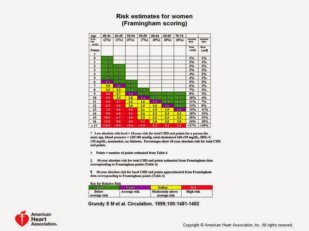
Risk of Cardiovascular Disease (by Framingham Scoring System)

Grundy et al. Circulation 1999;100:1481-82.

(Click on the image to enlarge)


8/30/2014

TEE - Basic Views & terminology

(From TEE for CHD Ed. by Wong PC & Miller-Hance WC. Springer-Verlag 2014)

(Click on the image to enlarge)


TEE - Probe position terminology

(Click on the image to enlarge)


Transesophageal echocardiography - Terminology of probe positions.
(From TEE for CHD. Ed. by Wong PC &Miller-Hance WC. Springer-Verlag 2014)

8/19/2014

VSD enlargement (Surgery)

To enlarge the VSD without causing AV block...

D-loop ventricles - Raise the roof (Enlarge VSD in anterior-superior direction)
L-loop ventricles - Lower the floor (Enlarge VSD in posterior-inferior direction)

Without injuring the aortic valve in the process!

7/28/2014

ICU: Postoperative AV block - Recovery

Study period: 1991-4. Boston Children's Hospital. n=2698 surgeries (73% on bypass).
Overall incidence of AV block was 3% (n=54).
Common diagnoses associated with development of AV block were VSD, Tetralogy, LVOTO repair, L-TGA and others. 
32 recovered conduction in 30 days after surgery (Graph below shows the postop. day at which conduction recovered in these 32 patients). 
9 did not recover conduction in 30 days after surgery. 
Weindling SN., et al. Duration of complete atrioventricular block after congenital heart surgery. Am J Cardiol 1998;82:525-527.


7/23/2014

ICU: Arterial Line Artifact - "Standing Wave"

Pardon the poor quality images.
Newborn, s/p Norwood procedure.
Upper panel shows arterial pressure traces from 2 different lines - Right radial arterial (Red) and Umbilical arterial lines (White). There is a "standing wave" or "fling" in the red trace. This is secondary to being a small/distal vessel, smaller catheter, and state of peripheral vascular tone (vasoconstricted state).

Lower panel shows the same patient, approx. 30 min after the loading dose of Milrinone. Presumably, there is adequate vasodilation of the peripheral artery that the "standing wave" or "fling" is no longer evident or not as prominent as before!

(Click on the images to enlarge)

4/28/2014

Echo artifacts

Five types of artifacts:
1) Mirror image artifact (Created by residual waves continuing to travel beyond the object of interest. Then, gets reflected back from interfaces beyond. These waves are once again reflected by the object giving rise to the second image.
 2) Reverberation artifact
 3) Side lobe artifact  (Produced by radial vibration of piezo electric crystals, instead of the longitudinal vibrations of the main beam)

4) Grating lobes artifact (no image) occurs in array transducers (?)

5) Acoustic shadowing (occurs due to highly reflective interface such as calcification).
(All images are from  TEE for congenital heart disease by Wong PC, Miller-Hance WC. Springer Verlog London. 2014.)